 |
|
|
|||||||||||||||||||||||||
|
| Nano and micro bio-engineering |
| Construction of bio-nano system using DNA Realization of tailor-made medical therapy |
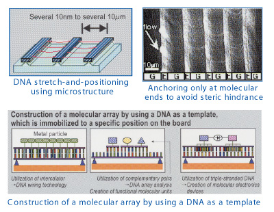
 Masao Washizu, Professor Masao Washizu, ProfessorDepartment of Mechanical Engineering DNA consists of four bases, A, T, G and C stacked at the spacing of 0.34 nm, and the genetic information is recorded by the sequence of the bases. DNA retains its structure, and reproduces itself, by the specific bindings between complementary bases, A-T and G-C. If a DNA strand is immobilized on a solid surface as a template, onto which the target molecules labeled with complementary DNA fragments are fed, the base pairs are spontaneously formed, and the target molecules will be aligned on the template as designed, at a resolution of 0.34 nm. Such a self-assembling technique will have applications in the construction of molecular electronics circuits, biochemical functional molecular units, or assembling of molecular machinery. The chemical stability of DNA, as well as the ease in its synthesis, makes the molecule the best candidate as the template for such molecular constructions. To investigate such possibility of DNA-based self-assembling technology, we are currently using our original electrokinetic DNA manipulation methodology in combination with microfabircation techniques to stretch and immobilize DNA strands in microstructures, and to perform dynamic interaction studies between DNA and foreign molecules. The clarification of the molecular process in single-molecule level will not only open a way for the molecular construction, but also provide basic knowledge for the improvement of hybridization-based gene analysis such as DNA chips. |
 |
| Development of Cell-Engineering Devices / Fujii |
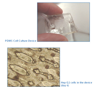
 Teruo Fujii, Associate Professor Teruo Fujii, Associate ProfessorDepartment of Environmental and Ocean Engineering The group of Prof. T. Fujii at the Institute of Industrial Science is working on the so-called 'Cell-engineering Devices' in collaboration with Prof. Y. Sakai's group at the Center for Disease Biology and Integrative Medicine. The idea of the device is to mimick the microfluidic environments in in vivo tissues by introducing microfabrication techniques. Three dimensional arrangement of the cells and active materials transport in the cultured tissues are expected to be realized in the newly developed devices. |
 |
| Real-time multipoint measurement of brain functions using ultra-precise electrodes: Development of auditory function recovering technology |
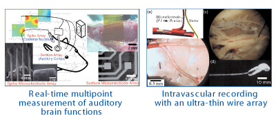
 Masayuki Nakao, Professor Masayuki Nakao, ProfessorDepartment of Engineering Synthesis The ultimate goals of our group are to elucidate the high-order function in the brain and to develop advanced medical technologies. Toward this purpose, we have been developing ultra microelectrode arrays on the basis of our microfabrication technologies, and have been applying them for mapping and microstimulation of the brain activities. The left figure shows, for instance, the animal model of neural prosthesis that directly stimulates the cochlear nucleus in the brainstem and restores hearing to deaf patients. Using the model, we mapped neuronal activities in the auditory cortex, and compared the activities evoked by acoustic stimulation and those by microstimulation in the cochlear nucleus. We developed a surface microelectrode array for the cortical recording and spike microelectrode array for the cochlear nuclear microstimulation. Our recent experiments highly suggest that microstimulation at an adequate location and that with adequate stimulation strength can induce the pitch and intensity sensation, respectively, thus substantially showing the capability of the prosthesis. The right figure(a) shows an intravascular recording with an ultra-thin wire array we are developing. In the recording, we introduce neural probes into cerebral vessels using a microcatheter and have the probes flow through capillaries toward the destination. The probes are required to introduce the probe to a spinal artery and measure the neural activities in the spinal cord. The right figure(d) shows the probe employing insulated platinum wire with a diameter of 1 mm, which was proved to be safe, and having platinum black electrodeposited at the tip, by which reduced electrical impedance yielded a sufficiently high S/N ratio. |
 |
Page top |